
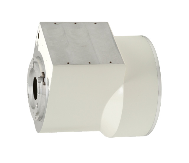
Electron Tubes & Devices E5764SD-P4A X-Ray Image Intensifier
X-ray image intensifier component
Price on request

Medical equipment and biomedical engineering.
Abuja, Nigeria.
Buy Canon image-intensifier fluoroscopy components in Nigeria through Franance by identifying the exact model and confirming compatibility with the installed C-arm or fluoroscopy system. The category lists E5764SD-P4A and E5830SD-P4A components; request a written quote order after technical review.
2 models
X-ray image intensifier component
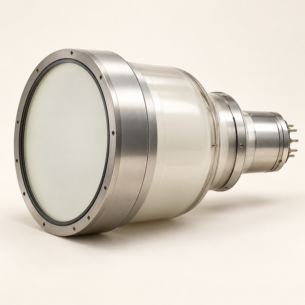
Request a quote order from Franance for one of the two Canon image-intensifier models currently listed: Canon E5764SD-P4A or Canon E5830SD-P4A. Start with the exact model code. Then provide the make, model, serial number and configuration of the fluoroscopy or mobile C-arm system in which the component is intended to be installed.
Read moreAn X-ray image intensifier is a component that converts the X-ray pattern passing through a patient into a corresponding visible-light image. Canon’s instructions for use describe the device as intended for installation in an X-ray fluoroscopy system and say that it does not itself record diagnostic images; a television camera is required to display the image on a monitor. That component-level definition matters when you compare quotations: the image intensifier alone is not the complete imaging chain.
Read moreThe Canon lineup lists both models in the medical 9-inch image-intensifier family. Both have a nominal normal-mode entrance field of at least 230 mm, a useful normal-mode field of at least 215 mm, M1 useful field of 160 ±5 mm, M2 useful field of 120 ±5 mm, and overall length of 338 ±5 mm. The listed output-image diameter is 20 ±0.5 mm for E5764SD-P4A and 25 ±0.5 mm for E5830SD-P4A.
Read moreBuy medical X-ray image intensifiers in Nigeria through Franance by identifying the exact replacement component, confirming compatibility with the installed fluoroscopy system, and requesting a written quote order. The two products currently assigned to this category are the Canon Electron Tubes & Devices E5764SD-P4A and E5830SD-P4A. Both are image-intensifier components listed for C-arm or fluoroscopy-table applications; neither is a complete C-arm, X-ray generator, camera, monitor or turnkey imaging room. Franance’s catalogue helps your team find the listed models and submit an enquiry. It does not take online payment or confirm stock, local approval, lead time or service coverage. Send the current system identity and component reference with your request. Franance can follow up by phone or email and confirm the actual scope and commercial terms in writing. A qualified technical reviewer should approve system compatibility before a replacement component is ordered.
Request a quote order from Franance for one of the two Canon image-intensifier models currently listed: Canon E5764SD-P4A or Canon E5830SD-P4A. Start with the exact model code. Then provide the make, model, serial number and configuration of the fluoroscopy or mobile C-arm system in which the component is intended to be installed.
An image intensifier is not a plug-and-play replacement simply because its field size or model family appears similar. The mechanical envelope, mounting surface, power supply, electrical connection, mode-selection circuit, optical lens and camera interface must match the host system design. Ask the system manufacturer or the qualified integrator responsible for the equipment to confirm the exact reference and revision. Franance can route the quotation enquiry and help clarify what information is needed; the catalogue assignment itself is not a compatibility statement.
The category sits under fluoroscopy and C-arm equipment. If you need a complete mobile system, compare mobile C-arm fluoroscopy systems; if the requirement is a room-based system, see fixed fluoroscopy systems. For a detector upgrade rather than an image-intensifier replacement, review flat-panel detector fluoroscopy. These are different scopes of supply.
An X-ray image intensifier is a component that converts the X-ray pattern passing through a patient into a corresponding visible-light image. Canon’s instructions for use describe the device as intended for installation in an X-ray fluoroscopy system and say that it does not itself record diagnostic images; a television camera is required to display the image on a monitor. That component-level definition matters when you compare quotations: the image intensifier alone is not the complete imaging chain.
This category currently contains two direct product records, both from Canon Electron Tubes & Devices. The E5764SD-P4A is listed with a 20 mm output-image diameter and is identified by the manufacturer for C-arm or fluoroscopy-table applications. The E5830SD-P4A is listed with a 25 mm output-image diameter and the same broad application description. Canon’s published specifications describe the shared 9-inch-class entrance fields and several performance figures, but those values do not prove either model will work in a particular system.
The category is deliberately narrower than C-arm systems. A mobile C-arm includes more than an intensifier: it is a configured X-ray imaging system with its own generator, controls, mechanical assembly and image chain. A fixed fluoroscopy room is likewise a larger system procurement. The two Canon catalogue records are components to evaluate for an existing compatible installation or a documented integration project. They should not be presented as complete systems or substituted for a full equipment schedule.
The Canon lineup lists both models in the medical 9-inch image-intensifier family. Both have a nominal normal-mode entrance field of at least 230 mm, a useful normal-mode field of at least 215 mm, M1 useful field of 160 ±5 mm, M2 useful field of 120 ±5 mm, and overall length of 338 ±5 mm. The listed output-image diameter is 20 ±0.5 mm for E5764SD-P4A and 25 ±0.5 mm for E5830SD-P4A.
Canon lists typical central resolution for E5764SD-P4A as 48, 56 and 66 lp/cm across normal, M1 and M2 modes; its typical contrast ratios are 25:1 over a 10% area and 16:1 over a 10 mm diameter. For E5830SD-P4A the corresponding typical resolution figures are 52, 58 and 68 lp/cm, with contrast ratios of 30:1 and 19:1. Canon lists 65% typical DQE for each. These figures are manufacturer-listed technical data, not a prediction of image quality in your installed system.
The E5830SD-P4A specification sheet additionally lists an approximate weight of 20 kg, a maximum diameter of 304 ±2 mm, and a 24 ±1 V DC input to its high-voltage power supply. Do not transfer those model-specific details to the E5764SD-P4A. Canon’s public lineup table shows the E5764SD-P4A’s mounting surface as the same side as the power-supply box and the front of the intensifier. Have the integrator check the drawing and exact assembly rather than using this summary as a dimensional approval.
These distinctions are useful for screening, not for choosing a replacement by headline numbers. Confirm the existing unit’s complete part number, system-generation compatibility, chosen input-field modes, output coupling and the integrator’s acceptance criteria. If the system documentation calls for a different output image size or mechanical arrangement, neither listed model should be assumed suitable. Request the manufacturer’s current document revision and a written compatibility decision for the actual host system.
Send the technical reviewer photographs of the nameplates and connections where permitted, the system make and model, serial number, installation configuration, existing intensifier part number and revision, and any applicable service documentation. Include the current camera, optical coupling or lens reference, power-supply identity, connector details and the exact reason for replacement. A service report or system drawing can help identify constraints, but it should be handled through the facility’s approved process.
Ask the responsible manufacturer or qualified integrator to confirm, in writing, at least the mechanical fit and mounting pattern; required orientation and clearance; electrical supply and high-voltage supply arrangement; mode switching and control connections; output image size and camera/optical interface; supported operating modes; and any system-specific software, calibration or adaptation work. Request the precise part number and revision on that confirmation. A nominal 9-inch family match does not settle any of these interface questions.
Also clarify who supplies and installs the component, who is responsible for integration, what documentation accompanies it, and what acceptance measurements are planned. If other parts of the image chain are to be replaced, identify each separately. The purchase order or quotation should state exclusions as well as inclusions so an intensifier component is not mistaken for an end-to-end fluoroscopy upgrade.
Start with the host system’s approved specification and the intended replacement reference, not a broad “image intensifier” description. Compare entrance field sizes and mode values, output image diameter, spatial-resolution figures, contrast-ratio data, DQE, mechanical dimensions, mounting surfaces and electrical requirements only where the exact source document supports them. Record units, test conditions, typical versus minimum values, document revision and whether an item is part of the quoted configuration.
Canon labels several values in its 9-inch lineup as typical. For E5764SD-P4A, the public table gives 48, 56 and 66 lp/cm central resolution for the listed normal and reduced modes, typical contrast ratios of 25:1 and 16:1, and 65% typical DQE. The E5830SD-P4A sheet reports 52, 58 and 68 lp/cm, 30:1 and 19:1 contrast ratios, and 65% typical DQE. These manufacturer measurements should be evaluated under their stated test conditions; they are not promises of diagnostic performance, patient outcomes or equivalence between installed systems.
The E5830SD-P4A technical sheet specifies overall length, maximum diameter, approximate weight and supply voltage. Check those alongside the system drawing and actual mounting conditions. For the E5764SD-P4A, use its exact model documentation or a written manufacturer response for dimensions or connections not fully stated in the lineup table. Never fill specification gaps by borrowing values from E5830SD-P4A or a related model.
Request the technical sheet and instructions for use that apply to the offered model and target country. Canon cautions that its product information may change and requires technical and regulatory verification before purchase and use. The available Canon IFU describes an EEA, Switzerland and Turkey objective area; it is not proof of Nigerian regulatory status or country eligibility. Ask Franance to identify the documents and terms actually applicable to the offered unit.
An image intensifier and a flat-panel detector are different detector technologies and component designs. This category contains two image-intensifier components; it does not include the Canon CXDI-RF Wireless B1 flat-panel detector, which has its own listing under flat-panel detector fluoroscopy. A detector component and an image-intensifier component should not be selected by broad modality label alone.
If your requirement is to replace a failed intensifier in an existing system, preserve the system’s exact part reference and obtain integrator approval for the proposed replacement. If the facility is evaluating a broader detector upgrade, compare full system compatibility, image-chain electronics, camera or detector interfaces, software, acquisition workstation, archive connectivity, installation work and acceptance scope. A detector technology change can involve system-level engineering and procurement; a category link does not establish that an upgrade is feasible.
If the institution is planning a new room or mobile unit, compare full fluoroscopy and C-arm systems, rather than treating an OEM component as a complete purchase. Define the required clinical service and approved equipment scope with the facility’s qualified team, then ask suppliers to respond against that requirement. The technical reviewers should decide whether a component replacement, system repair or complete system procurement is appropriate.
Send Franance the exact Canon model, required quantity, delivery facility, city and state, and the host fluoroscopy-system model and serial number. Attach the approved technical specification or component reference and list the interface information already confirmed by the manufacturer or integrator. If those details have not been confirmed, state that compatibility review is a requirement of the enquiry rather than assuming the part is suitable.
Ask the written quotation to identify the exact manufacturer entity, component model and revision, included documentation, configuration, exclusions, price, destination-specific delivery terms, lead time, warranty conditions and any installation or integration scope. Request separate line items for technical assessment, removal or replacement work, testing, acceptance, training, maintenance and other services if needed. Availability, local regulatory documentation, warranty, service coverage and delivery dates must be confirmed for the actual offer; this guide does not establish them.
For a tender, state whether the quote should be aligned to a named brand/model or a performance specification, and provide any required submission format and due date. Include your procurement contact and the qualified technical reviewer. Franance can follow up by phone or email to clarify the request. There is no online checkout: the enquiry is a quote order, and the written offer and subsequent agreement establish commercial terms.
An image intensifier is installed within a radiation-producing X-ray system. Component supply alone does not verify the performance or safety of the assembled system. Before work begins, agree with the system manufacturer or qualified integrator who is responsible for installation, electrical checks, mechanical checks, calibration, image-chain testing, documentation and system release. The facility should use its authorized engineering and radiation-safety procedures and applicable requirements.
IAEA Safety Guide SSG-46 describes acceptance testing and commissioning for medical radiological equipment. It says acceptance testing should verify specifications and features and be followed by commissioning; for fluoroscopy, a medical physicist should be directly involved in relevant measurements, calculations and interpretation. Use the facility’s qualified professionals to define the test plan and baseline for subsequent quality-control checks. This guide does not provide exposure settings, clinical protocols, shielding calculations or instructions to carry out technical service work.
Before returning a system to clinical use after a replacement, follow the applicable manufacturer procedures and facility approvals. Preserve records of the component identity, installation, tests, deviations and corrective actions. If the system experiences a defect that may affect safety, follow the manufacturer’s and facility’s procedures and keep it out of use as required by those procedures until qualified review is complete. The actual responsibilities and acceptance conditions should appear in the project agreement.
Send the destination city and state so Franance can check the proposed offer for that location. The catalogue is national in scope, but it does not promise that a particular model is available, deliverable or serviceable in every state. Confirm delivery, installation responsibilities, transport conditions, warranty, documentation, technical support and any market-specific requirements in writing for the actual transaction.
If one facility has multiple systems or locations, list each host system, component reference, quantity and destination separately. A single category selection does not confirm that the same model will fit every system. Keep technical compatibility and commercial availability as separate checks in the procurement file.
No. Franance does not accept online payment for these catalogue items. Submit a quote-order enquiry with the model and system details; the team will contact you by phone or email to clarify the requirement and prepare a written offer. Confirm technical fit with the responsible integrator and review the final commercial and regulatory documents before proceeding.
To start, send Franance the E5764SD-P4A enquiry or E5830SD-P4A enquiry, together with the host system make, model, serial number, existing component code, quantity, facility and destination. Franance can then follow up on a quotation for the specific requirement.
No. They are image-intensifier components. Each requires integration into a compatible X-ray system and image chain; the host system, camera, monitor, generator and other equipment are not included by category assignment.
Canon lists E5764SD-P4A with a 20 ±0.5 mm output-image diameter. E5830SD-P4A is listed with 25 ±0.5 mm. Confirm the required size and all interfaces against the exact host system documentation before ordering.
No. Check the full part number and revision, mounting geometry, electrical and mode-control interfaces, power supply, camera/optical coupling and system-specific acceptance requirements with the manufacturer or qualified integrator.
No. Manufacturer technical information does not establish current Nigerian availability, price, country eligibility, registration, delivery or service. Ask Franance to confirm the specific offer and documents in writing.
No. Submit a quote-order enquiry. Franance follows up by phone or email and confirms the commercial offer in writing.
Provide the exact component model, host fluoroscopy-system make, model and serial number, existing part reference, integrator compatibility confirmation, quantity, facility, city and state, and requested integration or acceptance scope. Franance will follow up to clarify the enquiry and confirm the offer and commercial terms in writing.
Request a quotationAdd the models you need to a quote order, or send us your equipment list.